These images come from GE's IN Cell Image Competition. The source article can be found here: http://www.newscientist.com/gallery/dn16308-inner-workings-of-cells/1
See comments below the images.
The trabecular meshwork is a key component of the eye, found at the base of the cornea (the transparent layer covering the front of the eye). It helps to drain liquid from between the cornea and the lens.
If the trabecular meshwork does not work properly, too much liquid builds up. The resulting increase in pressure is the number one treatable risk factor for glaucoma. Glaucoma is an optic neuropathy, which can lead to permanent loss of vision, which may progress to complete blindness if left untreated.
Many scientists are trying to understand the trabecular meshwork, in the hope of finding better treatments for glaucoma.
These cells are from the trabecular meshwork of a pig. Blue and green stains show, respectively, actin and tubulin - two key proteins that help maintain the shape of the cells and their interconnections. Red dots show focal adhesions, which are assemblies on the cells' outer membranes that allow them to interact with their surroundings.

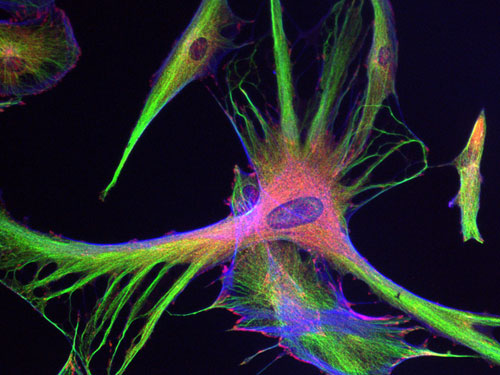
This close-up shows the internal structure of a single trabecular meshwork cell.
In this image, DNA has been stained blue, so the large clumps of blue just above centre are the cell's nucleus. Red lines are filaments of actin spread throughout the cell, while the green patches at their tips are the focal adhesions.
Filed Under (tags):
- dave's blog
- Log in or register to post comments
